
Case Study: TMEM42 and Brain Cancer

Image Credit: U.S National Library of Medicine
By: Jasmin Sangh
This case study explores the role of the TMEM42 gene and its relation to brain cancer. Through bioinformatic data and a fictional case study, this study highlights how abnormal TMEM42 expression may contribute to uncontrolled cell growth and tumor suppression. This study emphasizes the importance of awareness and continued research on TMEM42.
Gene Background
| Gene Name | Transmembrane Protein 42 |
|---|---|
| Gene ID | 131616 |
| Chromosome location | Chromosome 3p21.31 |
| Gene Type | Protein coding |
| Gene Function | Cellular stress response, nerve cell maintenance, immune regulation |
| Protein Type and Location | Multi-pass transmembrane- crosses the membrane more than once |
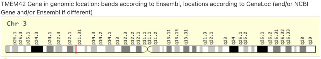
Figure 1: Genes Location– This figure indicates the genes exact location in the human body. The gene is located on chromosome 3 and is expressed in many parts of the body [1].
Bioinformatics Findings
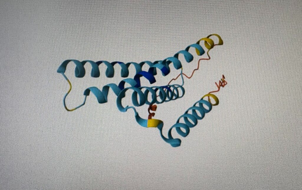
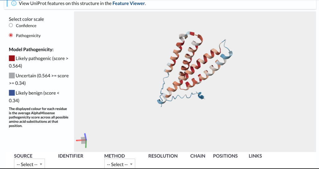
Figure 2a. Predicted 3D Structure of Transmembrane Protein 42– The protein is composed of alpha helices in a coiled coil structure. The dark and light blue segments on the protein structure indicate high confidence and the yellow and red structures indicate low confidence.
Figure 2b. Predicted 3D Structure of Mutated Transmembrane Protein 42– The end segment of the mutated protein binds to other sites in the cell and spreads the pathogens. Parts of the protein that are likely pathogenic are located in both dark and light red [2].
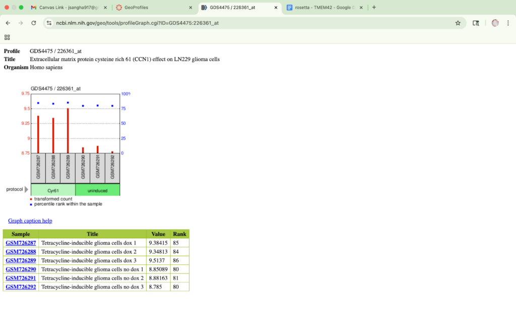
Figure 3. Cyr61 Induces TMEM42 Expression in Glioma Cells– Two groups are displayed in the figure and the red bars indicate TMEM42’s expression levels across both charts. TMEM42’s expression level increases in response to Cyr61 treatment. Cyr61 plays a role in the progression of tumorous gliomas by overexpressing itself [3][4].
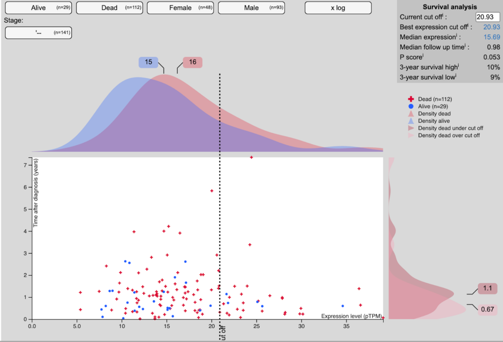
Figure 4. High TMEM42 Expression Correlates with Decreased Survival in Glioblastoma Patients– The data shows that patients with higher TMEM42 expression have lower survival rates than those with lower expression. The average survival of a patient after three years with high expression is 9% as opposed to 10% for lower expression. The red crosses on the scatter plot indicate the number of individual dead patients and the blue dots show the number of alive [5].
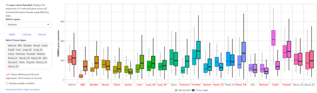
Figure 5. TMEM42 Gene Expression in Body Cells– The graph indicates the gene’s expression in normal and tumorous cells. The first box plot shows the gene’s normal expression in the bladder, breast, stomach, and other cells. The second plot shows the expression after the cell becomes tumorous [6].
Associated Phenotype/Disease
Mutations in the TMEM42 gene seem to be linked to a higher risk of brain tumors; specifically types like glioblastoma. TMEM42 encodes a transmembrane protein found in the cell membrane, where it likely helps regulate cell communication and growth. When mutated, TMEM42 can produce an abnormal transmembrane 42 protein that disrupts normal cell function, leading to unregulated cell division. This uncontrolled growth can help contribute to the formation of brain tumors and neurodegenerative diseases. A specific mutation in the TMEM42 gene where c.445C>T is found in the glioma cells. TMEM42 is expressed in glioma cells and patient tumor samples. This shows its important role in brain cancer development. With this being said, no clinical relation has been documented between TMEM42 and Glioma cells. Other transmembrane gene family members have been linked to be a prognostic factor and have been found to be linked with aggressive brain tumors and other neurodegenerative diseases [7].
Fictional Case Study
A 52-year-old female began having severe headaches and memory lapses. After getting an MRI, it was revealed that she had a brain tumor in her left temporal lobe, later diagnosed as a glioblastoma multiforme (GBM). Genetic testing of the tumor showed overexpression of the TMEM42 gene, which is known to be active in glioma cells and associated with other tumor behavior and neurodegenerative diseases. Due to the tumor’s location and fast growth, the female went through surgery followed by radiation and chemotherapy.
References
- UCSC Genome Browser. (2025). Human hg38 chr3:44,861,904-44,865,670 UCSC Genome Browser v485. Ucsc.edu. https://genome.ucsc.edu/cgi-bin/hgTracks?db=hg38&lastVirtModeType=default&lastVirtModeExtraState=&virtModeType=default&virtMode=0&nonVirtPosition=&position=chr3%3A44861904%2D44865670&hgsid=2866225940_gpHQJTlhzzqyjqA9aaXYgcIbap9p
- Uniprot. (2025). UniProt. UniProt. https://www.uniprot.org/uniprotkb/Q69YG 0/entry
- NCBI Gene Profile. (2024). transmembrane protein 42. NCBI. https://www.ncbi.nlm.nih.gov/datasets/gene/131616
- NCBI. (n.d.). CCN1 cellular communication network factor 1 [Homo sapiens (human)] – Gene – NCBI. Www.ncbi.nlm.nih.gov. https://www.ncbi.nlm.nih.gov/gene/3491
- The Human Protein Atlas. (2021). Brain tissue expression of TMEM42 – Summary. Proteinatlas.org. https://www.proteinatlas.org/ENSG00000169964-TMEM42/brain
- TNMplot. (2013). TNMplot: differential gene expression analysis in Tumor, Normal and Metastatic tissues. Tnmplot.com. https://tnmplot.com/analysis/
- Li, B., Huang, M.-Z., Wang, X.-Q., Tao, B.-B., Zhong, J., Wang, X.-H., Zhang, W.-C., & Li, S.-T. (2015). TMEM140 is associated with the prognosis of glioma by promoting cell viability and invasion. Journal of Hematology & Oncology, 8(1). https://doi.org/10.1186/s13045-015-0187-4